Heart block
Heart block (ATRIOVENTRICULAR (AV) BLOCK)
In this condition conduction between the atria and ventricle is impaired.
There are three forms of AV heart block
1 . First-degree heart block
2, Second degree (partial) heart block
3- Third-degree (complete) heart block
FIRST-DEGREE HEART BLOCK
It is a simple prolongation of the PR interval to more than 0.22 seconds.
Every impulse from the atria is conducted to the ventricle but with delay.
First-degree heart block implies prolonged conduction without block, each atrial impulse is conducted to the ventricles but there is a delay in AV conduction leading to prolonged PR interval on ECG.
Site:– The site of slowed conduction may be within the atrium, AV node, bundle of His, or bundle branches.
Causes
Enhanced vagal stimulation such as in inferior wall MI
Drugs e.g, digoxin, beta-blockers, verapamil, diltiazem and amiodarone.
Myocarditis, Addison disease
Congenital heart diseases such as ASD
Rheumatic fever
It can also occur in normal individuals such as children and athletes.
Clinical features
It cannot be diagnosed clinically and its recognition depends on observing a PR interval of >0.20 s in ECG.
Management
It is a benign condition and requires no treatment
Its only importance is as an index of drug toxicity and as a precursor of the more advanced degrees of heart block.
Second-degree heart block
Second-degree heart block implies intermittent conduction; some impulses from the atria are conducted to ventricles whereas others are not.
There are 2 main types of second-degree heart block
Mobitz type 1
In this condition, there is a progressive prolongation of successive PR intervals followed by a dropped beat (non conducted P wave). This is also known as Wenckebach’s phenomenon. In this AV block, there is a conduction defect in the AV node and AV conduction time (PR interval) progressively lengthens before the blocked beat. Pulse is irregular clinically.
Prognosis is good in first-degree and in Mobitz type 1.
The site of the block is mainly an AV node. QR$ complex is normal in morphology (not wide) because there is no delay in intraventricular depolarization.
Causes
Inferior wall MI
Acute rheumatic fever
Myocarditis
Degenerative conductive system disease.
Drugs such as digoxin, beta-blockers,s and calcium channel blockers
Hyperkalemia
Mobitz type 2
In this condition, the PR interval of the conduct impulses remains constant but some P waves are not conducted which is more P waves than QRS complex.
The risk of complete heart block in this type is greater than Mobitz type 1. The site of the block is intranodal in location and the QRS complexes are wide
Mobitz type II AV block is abrupt and is not preceded by lengthening of AV conduction time. It is usually due to blocking within the bundle of His.
Can lead to complete heart block
Management
Treat the cause
Atropine if needed
Pacing may be required
COMPLETE HEART BLOCK
A complete heart block is an advanced form of block. No impulse from the atria reaches the ventricles. Cardiac action is maintained by an escape rhythm.
Escape rhythm arising in the bundle of His produces narrow QRS complexes at the rate of 50-60 beats per minute. Escape rhythm arising below the His bundle produces broad complexes and at the rate of 15-40 beats/min.
Exercise does not increase the heart rate
ETIOLOGY OF COMPLETE HEART BLOCK
Congenital
Acquired
Idiopathic fibrosis
Myocardial infarction/ischemia
Infections, aortic root abscess in infective endocarditis
Chaga’s disease
Lyme disease
Sarcoidosis
Amyloidosis,
Neoplasia
Trauma e.g. cardiac surgery
Drugs e.g. digoxin, beta-blockers, amiodarone
Connective tissue disease: SLE, Rheumatoid arthritis
SYMPTOMS OF HEART BLOCK
Symptoms of heart block develop due to bradycardia
Exercise intolerance
Easy fatigability
Dyspnea on exertion
Syncope
Dizziness
MANAGEMENT
Acute inferior wall myocardial infarction is often complicated by transient AV block which may respond to atropine. if it fails temporary pacemaker should be inserted. In many cases, the AV block will resolve within 7-10 days. The patient may develop heart failure or hypotension due to a slow heart rate in an already damaged heart requiring a pacemaker.
Mobilz II or complete heart block as a result of anterior wall infarction is usually a sign of extensive myocardial damage and carries a poor prognosis.
Asystole often occurs and a temporary pacemaker should be inserted prophylactically as soon as possible.
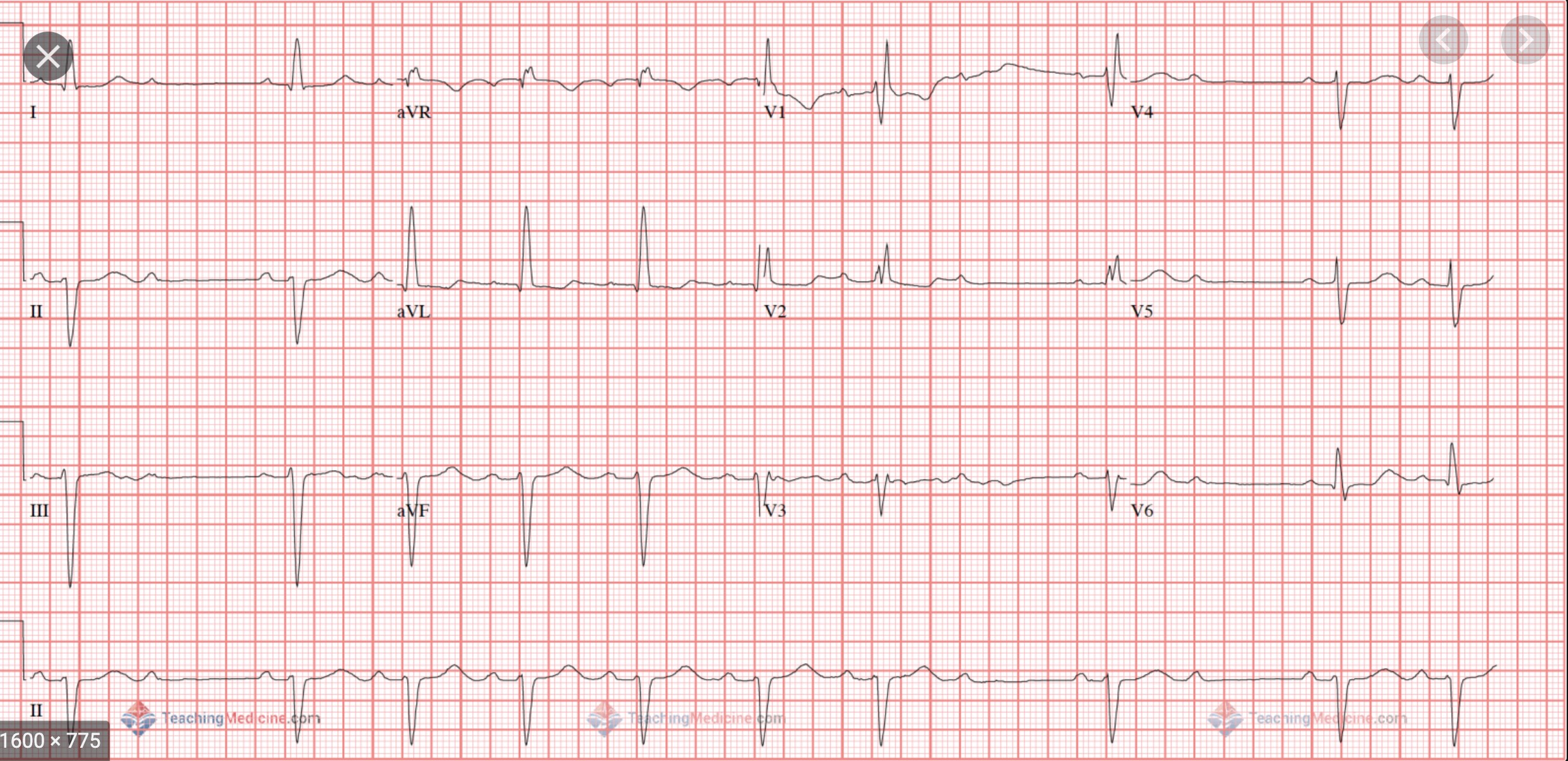
2nd-degree heart block- Mobitx type-1


2nd-degree heart block- Mobitx type-2